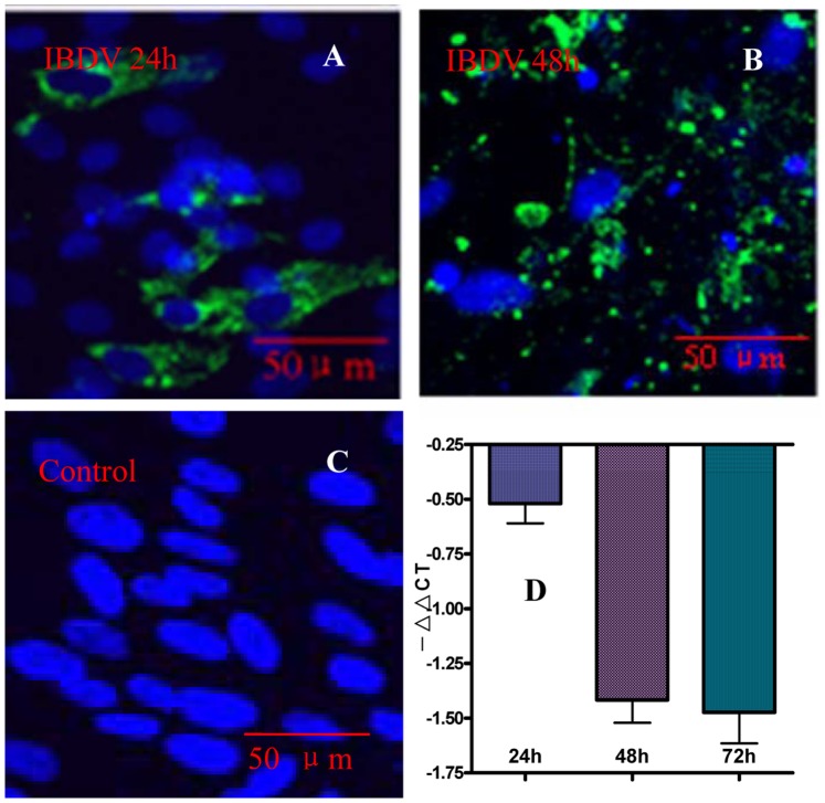
Figure 2. The mRNA abundance and protein expression of chCD132 on an IBDV-infected CEF monolayer.
CEFs were infected with IBDV a 100 TCID dose of the eNB virus. (A)–(C) Double-stained immunofluorescence images with anti-chCD132 mAb (red) and chicken serum (green) to IBDV under laser confocal microscopy. (A), (B) and (C) There is no chCD132 (red) expression in IBDV- and mock-infected CEF. (D) Transcription kinetics of chCD132 analyzed by qRT-PCR. Samples were normalized with the β-actin gene as a control and uninfected CEF at each time point as a reference. Each experiment was conducted in triplicate. Values are expressed as −ΔΔCT ± SD.

