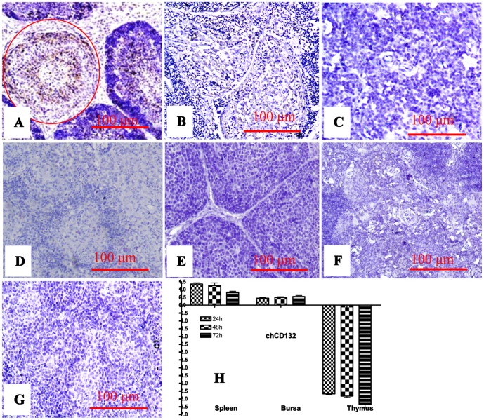
Figure 3. Expression and transcription of chCD132 mRNAs in SPF chickens infected with IBDV.
(A)–(G) represents immunohistochemical staining of bursa, thymus, and spleen of mock- and IBDV-infected chickens. (A) IBDV antigens (brown) in bursa lymph follicles (red, round); (B) Bursa lymph follicles of IBDV-infected chicken unrecognized by anti-chCD132 mAb; (C) Thymus of IBDV-infected chicken unrecognized by anti-chCD132 mAb; (D) ChCD132 antigens in spleen of IBDV-infected chicken unrecognized by anti-chCD132 mAb; (E)–(G) Bursa, thymus and spleen of mock-infected chickens un-reactive with anti-chCD132 mAb; (H) chCD132 mRNA transcription in bursa, thymus, and spleen of chicken infected with IBDV. Samples were normalized with the β-actin gene as a negative control, and uninfected SPF chicken as a reference. All samples were assayed in triplicate and the values are expressed as −ΔΔCT ± SD.

