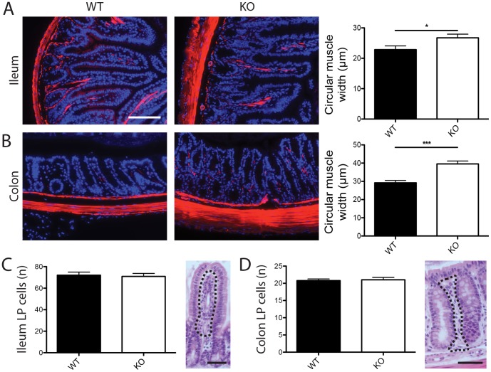
Figure 5. Intestinal muscle width is increased in TM-IEC C1galt -/- mice in absence of immune cell infiltration.
(A–B) Smooth muscle actin stainings and quantification of sections from ileal (A) and colonic (B) tissue of wild type (WT) and TM-IEC C1galt -/- (KO) mice. (C–D) Quantification and representative image of ileal (C) and colonic (D) lamina propria cells. LP - lamina propria. The dotted lines represent the areas used for scoring. For quantifications n = 5–7 mice were scored. Scale bars indicate 50 µm. Data shows mean ± SEM; * p<0.05, *** p<0.001.

