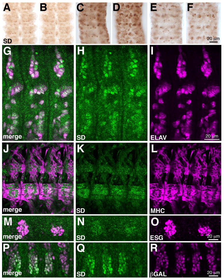
Figure 3. SD expression in the embryonic CNS, PNS, limb primordia, and somatic muscles.
(A–F) Expression of SD expression VNCs dissected from stage 12 (A, B), stage 15 (C, D) and stage 16 (E, F) embryos. For each stage, dorsal (A, C, E) and ventral (B, D, F) focal planes are shown. SD is initially broadly expressed in the VNC, and expression resolves to subsets of cells as development proceeds. (G–I) SD (green) and ELAV (magenta) in three abdominal segments of a stage 12 embryo, showing that most SD expressing cells are sensory neurons. These images are maximum projections of multiple confocal slices. (J–L) Four abdominal segments of a late stage 16 embryo showing SD (green) expression in muscles VL1–4 and LL1, which are marked by MHC (magenta). (M–O) SD (green) is coexpressed with ESG (magenta) in the dorsal limb primordia of the second and third thoracic segments of a stage 17 embryo. These images are maximum projections of multiple confocal slices. (P–O) Three thoracic segments and first abdominal segment of a stage 12 embryo. SD (green) is coexpressed with βGAL driven by twist-GAL4 in the three thoracic segments. Specimens are oriented with anterior up (A–F) or to the left (G–R).

