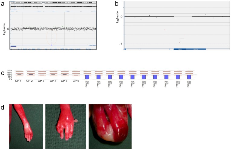
Figure 2. Results from array-CGH and MLPA analysis on genomic DNA from fetal case FC10.
Upper images show a hemizygous deletion (mean log2-ratio -2.5) on Xp22.2 comprising two probes located within the FANCB gene consistent with a diagnosis of X-linked VACTERL (a, b). Results from MLPA analysis of FANCB in fetal case FC10 (c). Y-axis scale with 100% representing a normal copy number and values above and below representing duplications and deletions respectively (0% representing homo-/hemizygous deletion). Results shown as blue bars emanating from baseline (100%). Analysis results in FC10 showing six control probes with normal copy number and hemizygous deletions of all FANCB exons. Lower images show malformations identified in fetal case FC10: left thumb agenesis (left), right thumb hypoplasia (middle) and imperforate anus (right) (d).

