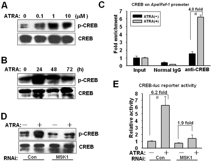
Figure 5. MSK1-mediated CREB phosphorylation activates Ape/Ref-1 expression.
(A) U266 cells were treated with the indicated concentrations of ATRA for 4 h. Cell lysates were subjected to immunoblotting with antibodies to phosphorylated CREB (Ser133) and total CREB. (B) U266 cells were incubated with 1 µM of ATRA for the indicated periods. Phosphorylated and total CREB levels were measured by Western blotting. The figure shows a representative result of triplicate experiments. (C) After exposure to 1 µM ATRA for 24 h, U266 cells were subjected to ChIP experiments using anti-CREB and control normal IgG antibodies. Immunoprecipitated genomic DNA fragments were amplified by PCR with specific primers targeting Ape/Ref-1 promoter. Input reflected the relative amounts of sonicated DNA fragments using in immunoprecipitation. ATRA vs. vehicle: # P<0.01, n = 3. (D) At 24 h after transfection of scrambled or MSK1 siRNAs, cells was treated with 1 µM of ATRA for another 24 h. Attenuated CREB phosphorylation by MSK1 knockdown was detected by Western blotting. (E) At 24 h after transfection of siRNAs against MSK1 together with CREB-luc (2 µg) and pRSV-luc (20 ng) reporters, U266 cells were incubated with 1 µM ATRA for another 24 h. Luciferase activity was measured and normalized to Renilla luciferase activity. Each value presented is the average of triplicate samples and a representative of multiple independent experiments.

