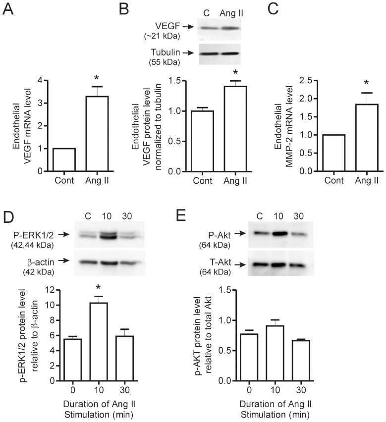
Figure 5. Effect of Ang II stimulation on VEGF and MMP-2 expression in skeletal muscle endothelial cells.
Skeletal muscle endothelial cells were stimulated for 2 hours with Ang II (0.1 µM) and lysed for qPCR analysis of VEGF (A) and MMP-2 (C) transcript levels (n = 6 per condition). In (B), endothelial cells were treated overnight with Ang II (0.1 µM) and Western blotting was performed to assess VEGF protein levels, which were normalized to tubulin (n = 3 per condition). Endothelial cells were stimulated for either 10 or 30 minutes with 1 µM Ang II and then lysed for protein analysis. P-ERK1/2 (D) and P-Akt (E) levels were assessed by Western blotting and normalized to ß-actin and total Akt levels respectively (n = 3 per condition). Values are presented as mean ± SEM. One way ANOVA followed by Tukey’s multiple comparison test and student’s t-test were used to assess statistical significance (p<0.05). In panels A–D, *denotes p<0.05 vs. untreated cells. C – Control, 10–10 minutes and 30–30 minutes.

