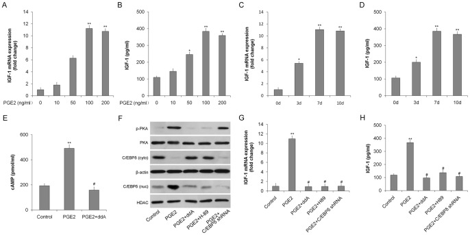
Figure 3. IGF-1 expression in rat TSCs is upregulated by PGE2 via the cAMP/PKA/CEBPδ pathway.
TSCs were incubated with 0, 10, 50, 100, or 200/ml PGE2 for 7 days. (A) IGF-1 mRNA expression was determined by qRT-PCR; (B) IGF-1 protein levels were quantitated by ELISA. TSCs were incubated with 100 ng/ml PGE2 for 0, 3, 7, or 10 days. (C) IGF-1 mRNA expression was determined by qRT-PCR; (D) IGF-1 protein levels were quantitated by ELISA. (E) TSCs were incubated in medium containing PGE2 (100 ng/ml), with or without the cAMP inhibitor ddA (10 μmol/l) and intracellular cAMP levels were determined by EIA. TSCs were incubated in medium containing PGE2 (100 ng/ml) with or without the cAMP inhibitor ddA (10 μmol/l), the PKA inhibitor H-89 (10 μM), or after transduction with the CEBPδ shRNA. (F) p-PKA and total PKA, nuclear (nuc) and cytoplasmic (cyto) CEBPδ protein levels determined by western blotting. HDAC (nuc) and β-actin (cyto) were used as loading controls; (G) IGF-1 mRNA levels determined by qRT-PCR. The mRNA levels were normalized using GAPDH; (H) IGF-1 protein levels determined by ELISA. Results represent the mean ± SD. *P<0.05, **P<0.01 with respect to TSCs without PGE2; #P<0.05 with respect to TSCs with PGE2.

