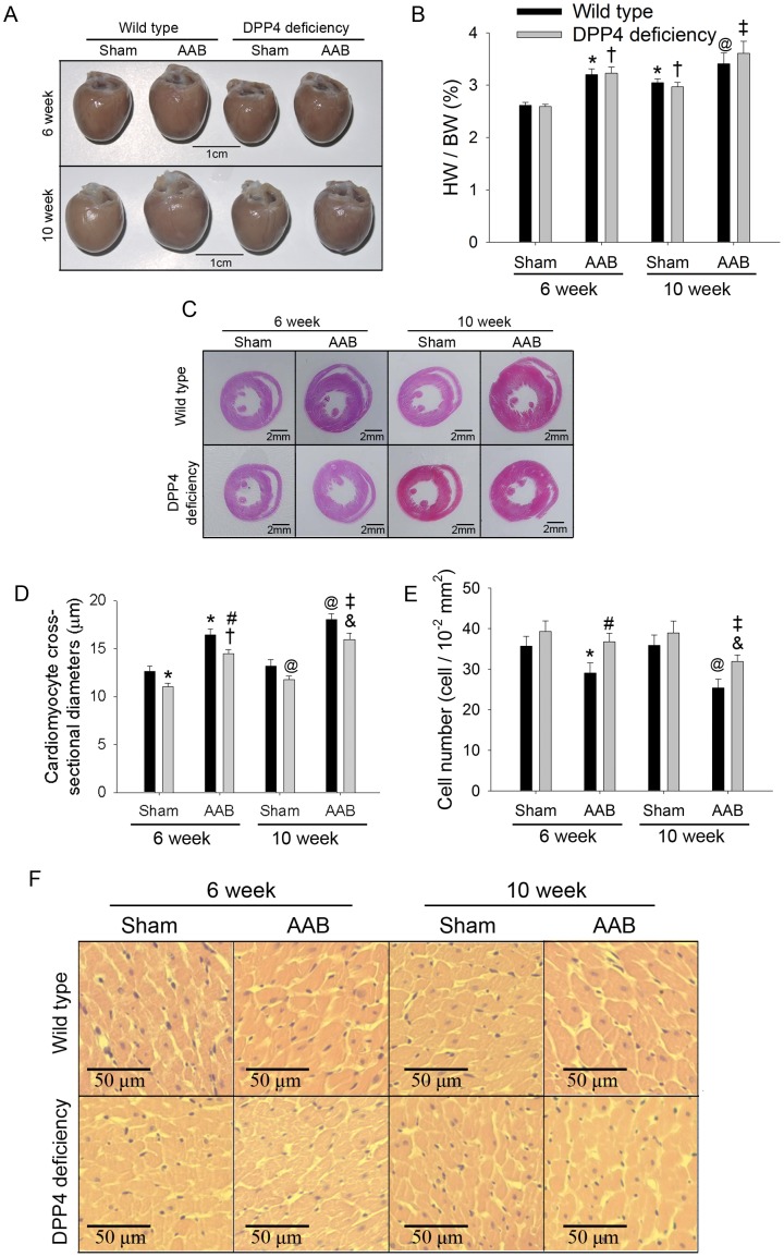
Figure 2. Effects of AAB on heart morphology.
Representative heart (A), heart weight to body weight ratio (n = 8) (B), and histological sections (C and F) of HE staining were shown after 6 and 10 weeks of AAB surgery. Cardiomyocyte cross-sectional diameter (D) and cell number (E) were also measured. (300 cell in 5 rats each). *p<0.05 vs. wild-type 6 week sham, #p<0.05 vs. wild-type 6 week AAB, @p<0.05 vs. wild-type 10 week sham, &p<0.05 vs. wild-type 10 week AAB, †p<0.05 vs. DPP4 deficiency 6 week sham, ‡p<0.05 vs. DPP4 deficiency 10 week sham.

