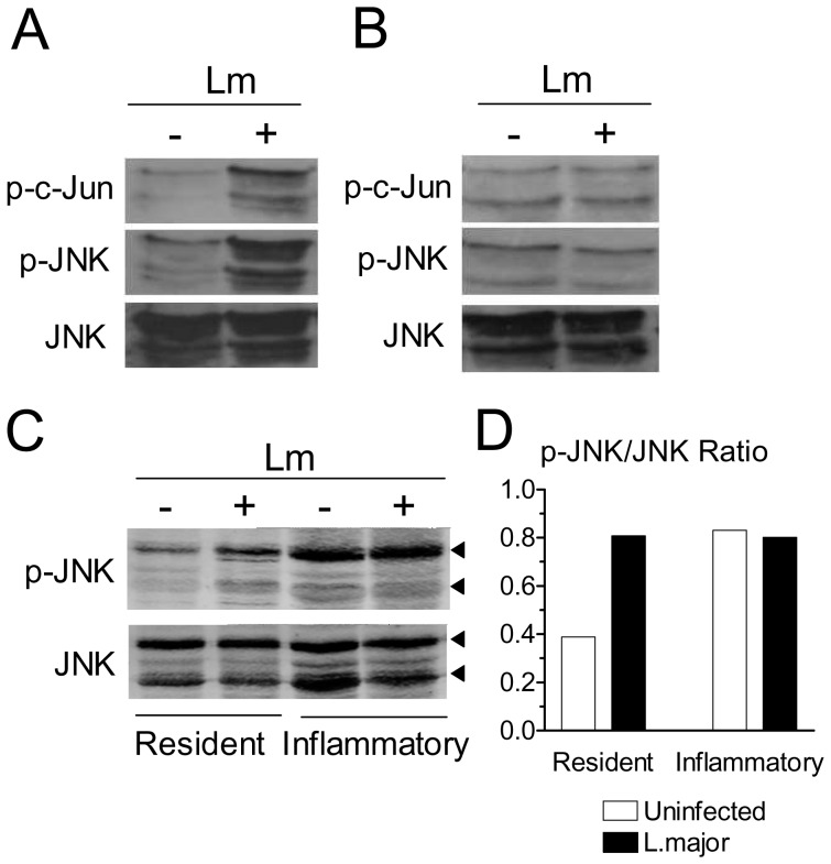
Figure 2. Infection with L. major activates the SAPK/JNK pathway.
Resident (A) or inflammatory (B) B6 adherent macrophages were infected or not with L. major (Lm). After 4 h, cell extracts were obtained and the levels of JNK, p-JNK and p-c-Jun were determined by western blotting. (C) Resident and inflammatory macrophages were adhered and infected in parallel. After 4 h, the levels of JNK and p-JNK were determined by western blotting. Top and bottom arrowheads indicate the p54 and p46 JNK bands, respectively. (D) Densitometric analysis of the blot shown in Figure 2C. The areas of both p46 and p54 bands were scanned. Results were normalized as the ratio between the intensities of the p-JNK and total JNK bands.

