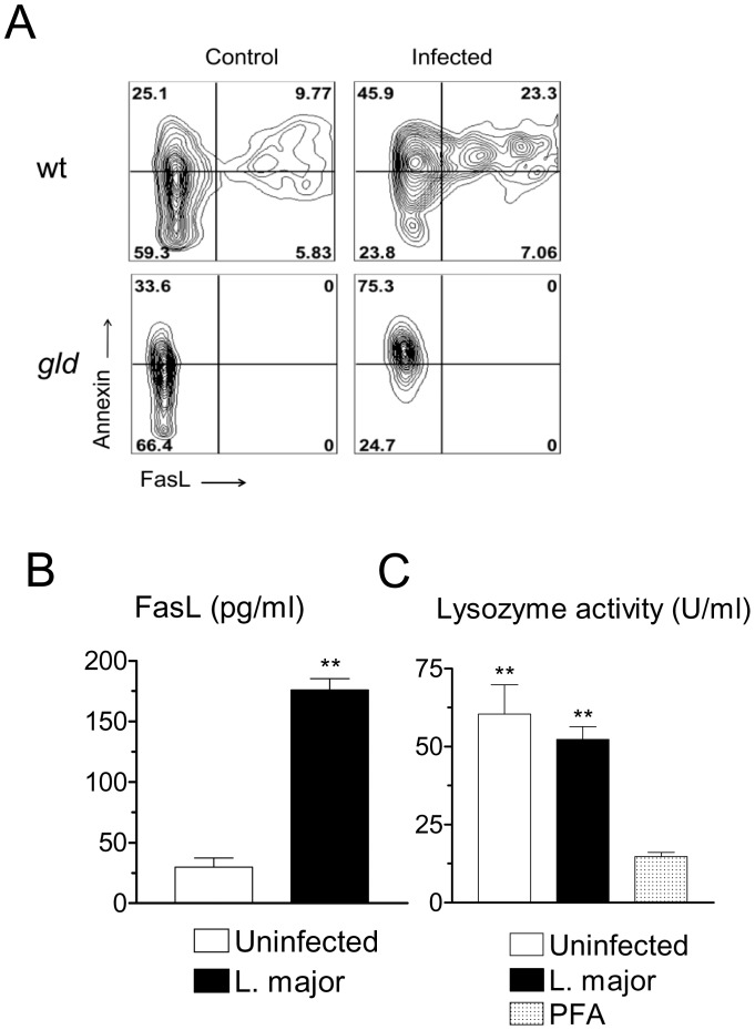
Figure 3. Infection with L. major increases FasL expression, but does not induce cell death.
(A) Resident B6 macrophages, either wild-type (wt) or FasL-deficient gld, were infected or not for 20 h. Monolayers were detached and stained for FCM. Gated populations comprise F4/80+ CD11b+7AAD− viable macrophages. Results indicate contour plots of Annexin V versus FasL staining. Numbers indicate percentages of cells in each quadrant. (B) Levels of soluble FasL in the supernatants of either control or infected resident macrophages 28 h after infection. (C) Resident macrophages were infected with L. major and cultured for 48 h. Supernatants were assayed for lyzozyme activity. As a control, macrophages were treated with paraformaldehyde (PFA) before collecting the supernatant. Results are mean and SE of triplicate cultures. **P<0.01.

