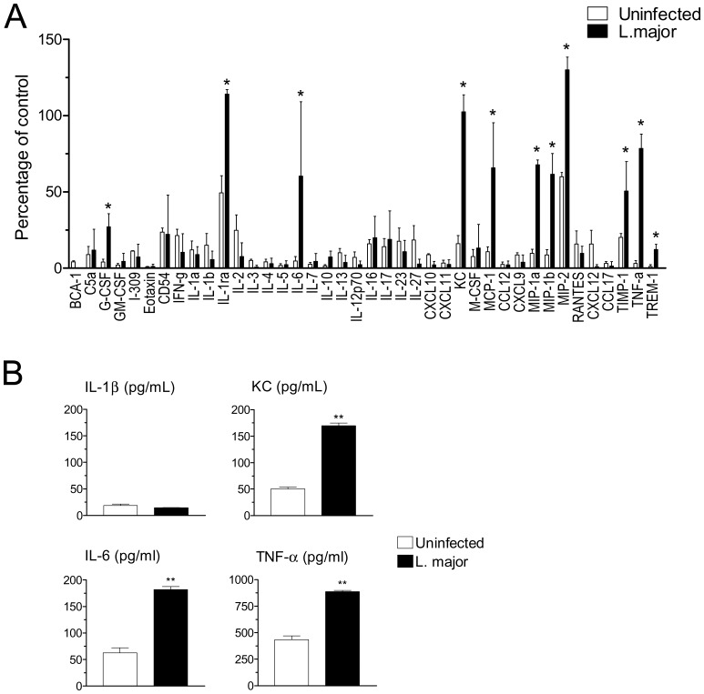
Figure 4. Induction of cytokine and chemokine release by L. major infection.
(A) Resident B6 macrophages were infected (closed bars) or not (open bars) for 20 h with L. major, and supernatants were probed with a mouse cytokine array. The intensity of the labeling for each cytokine/chemokine/mediator was quantitated and normalized as percentage of a positive control provided in the kit. Data indicate mean and SD of two independent arrays. Infected versus uninfected values were compared using non-parametric Mann-Whitney U-test. Cytokines showing a significant (P<0.05) increase following infection are indicated by an asterisk. (B) Supernatants were also probed by ELISA for IL-1β, KC, IL-6 and TNF-α, as indicated. **P<0.01.

