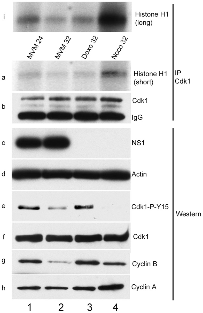
Figure 4. Inhibitory phosphorylation of CDK1 is transient during infection even though cells remain blocked in G2.
Para-synchronized A9 cells were infected for the indicated time points. Control cells were treated with 200 nM doxorubicin (doxo, lane 3) or 150 ng/ml of nocodazole (noco, lane 4) 16 hours post release (just after S-phase entry) and harvested at the indicated time points. Cells were lysed in IP kinase buffer as described in materials and methods. Equal amount of lysates were immunoprecipitated with 4 µg of CDK1 antibody and used for kinase assays using histone H1 as substrate. Panel b shows amounts of immunoprecipitated CDK1. Autoradiograms of phosphorylated histone H1 are shown in panels a (short exposure) and i (long exposure). Input samples were blotted with antibodies directed against NS1 (panel c), actin (panel d), total CDK1 (panel f), CDK1 phosphorylated on tyrosine 15 (CDK1-P-Y15, panel e) and cyclins A (panel h) and B (panel g).

