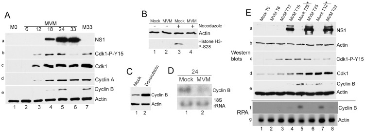
Figure 5. Virus-mediated downregulation of cyclin B1 RNA prevents mitotic entry.
(A) Cyclin B1 loss late in MVM infection. Time course of MVM infection of para-synchronized A9 cells. Mock cells were harvested at the time of release (M0, lane 1) or 33 hours post release (M33, lane 7). Western blotting was performed as described in Figure 4, using antibodies directed against NS1 (panel a), CDK1-P-Y15 (panel b), total CDK1 (panel c), cyclin A (panel d), and cyclin B1 and actin (panel e). (B) Infected cells do not proceed into mitosis. Nocodazole trap assay was performed as described in Materials and Methods. Cells were blotted using antibodies against actin and histone H3 phosphorylated on serine 28 (Histone H3-P-S28) (C) Doxorubicin treatment leads to cyclin B1 accumulation. Mock treated and 200 nM doxorubicin treated cells were harvested at 24 hours post treatment and blotted using antibodies directed against cyclin B1 and actin. (D) MVM infection results in reduction of cyclin B1 RNA at 24 hpi. Northern blot analyses comparing RNA from parasynchronized mock and MVM infected cells at 24 hpi. Ethidium bromide stained 18s rRNA served as a loading control. (E) Time course analyses comparing RNA and protein from mock and infected cells late in infection. Samples harvested at the indicated time points were processed for western blotting (panels a to e, using antibodies described in Fig. 4A), and RNAse protection assays (RPA, panels f and g) using probes against murine actin and cyclin B1. * - nocodazole added at 19 hpr.

