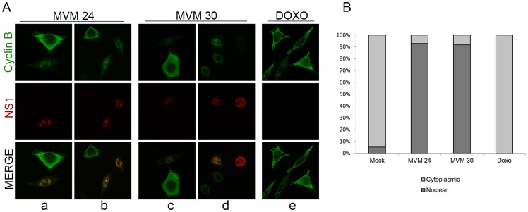
Figure 6. MVM infection induces premature nuclear entry, recruitment into APAR bodies and eventual loss of cyclin B1.
(A) Mislocalization of cyclin B1. Panels a to d; para-synchronized A9 cells were infected with MVMp (MOI of 10) for 24 or 30 hours before being fixed and processed for immunofluorescence. APAR bodies were detected with antibodies to NS1. Representative images are shown. G2 cells (identified by prominent cytoplasmic staining of cyclin B1) are shown juxtaposed to infected cells. In infected cells which showed cyclin B1 staining, cyclin B1 was nuclear and observed within distinct foci which co-localized with APAR bodies. Also, cyclin B1 staining intensity was reduced in many infected cells (compared to G2 cells, see figure 6C) and was completely absent in some (see Figure 6D, compare infected cell on the far right with the one adjacent). All images were captured using an objective of 63×. Panel e; A9 cells were treated with 200 nM doxorubicin (doxo) for 28 hours and processed as above. (B) Quantitation of the intracellular distribution of cyclin B1. Mock, doxorubicin-treated and MVM infected A9 cells from the experiment in (A) and an additional experiment were quantified to indicate the cellular region showing predominant cyclin B1 staining. Approximately 100 cells with detectable cyclin B1, in 7 randomly selected fields, were scored in an unbiased manner and plotted. Mock cells in G0/G1 as well as MVM-infected cells with significantly reduced cyclin B1 staining were not included in the scoring.

