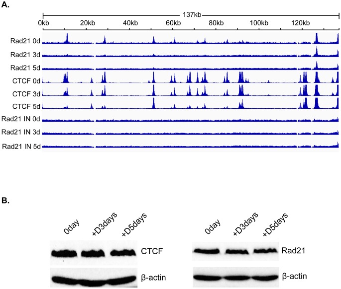
Figure 5. Changes in Rad21 binding to the KSHV genome during KSHV reactivation and lytic replication.
A. ChIP-SEQ assays were performed on cell samples harvested at 0 d, 3 d, and 5 d after induction of lytic KSHV replication in iSLK cells. Relative read numbers are plotted on the vertical axis versus the reference KSHV genome map on the horizontal axis. Rad21 ChIP results from each time point are shown on the upper three panels and the corresponding CTCF ChIP results are shown on the middle three panels for comparison. The corresponding input samples (IN) are shown on the lower three panels. The tracks depict coverage per base, scaled per million mapped KSHV reads. B. CTCF and Rad21 levels in iSLK cells during KSHV replication. Protein lysates from iSLK cells at 72 and 120 hours after induction of lytic replication were immunoblotted with anti-CTCF or Rad21 antibodies. Blots were stripped and re-probed with anti-actin antibody as a loading control (lower panels).

