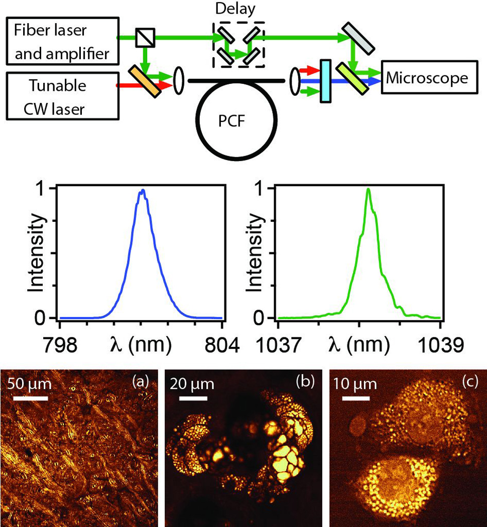
Figure 5. Fiber source for CARS microscopy and imaging of mouse tissue.

Top: schematic of fiber source based on FWM in PCF. Middle: spectra of generated pulses. Bottom: CARS images at the 2850 cm−1 mode of CH2: (a) mouse brain, (b) sebaceous gland 40 µm deep in mouse ear and (c) isolated rat fibroblast cells. From Ref. 79.
