
FIGURE 2.
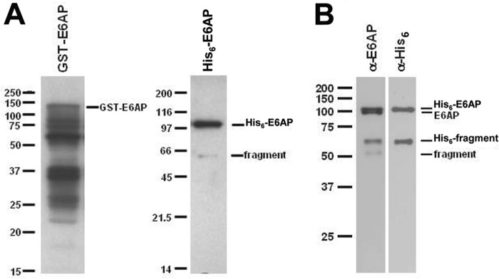
SDS-PAGE of selected E6AP preparations. A, Coomassie-stained 10% (w/v) SDS-PAGE resolution of affinity-purified recombinant GST-E6AP expressed in E. coli (left) versus His6-E6AP expressed in baculovirus (right). B, Western blot of 12% (w/v) SDS-PAGE resolution of baculovirus-expressed His6-E6AP stained with anti-E6AP antibody (left) and then stripped and restrained with anti-His6 antibody (right). Mobility markers are shown to the left of the corresponding panels. Mobilities of selected E6AP species are shown to the right of the corresponding panels.