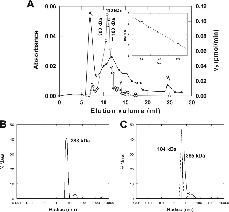
FIGURE 3.
Functional E6AP is an oligomer. A, 150 μl of 75 μm full-length His6-E6AP was analyzed in a 1× 30-cm Superose 12 FPLC gel filtration column equilibrated in 50 mm Tris-HCl (pH 7.5) and 50 mm NaCl. Protein was monitored by 280 nm absorbance (filled circles), and enzyme activity was monitored by E3-limiting 125I-polyubiquitin chain formation (open circles). Inset, calibration plot with the elution position of the peak E6AP activity shown with an open circle. B, static light scattering analysis of 18 μm full-length His6-E6AP in 50 mm Tris-HCl (pH 7.5) containing 200 mm NaCl at 37 °C as described under “Materials and Methods.” The main peak of 283 kDa exhibits a polydispersity of 17%. The higher molecular weight low abundance peak of 50-nm radius represents residual aggregates not removed by Mono Q FPLC. C, static light scattering analysis identical to A in the presence of 16 μm His6-E6AP and 8% (v/v) methanol in the absence (solid line; 23% polydispersity) or presence (dashed line; 14% polydispersity) of 61 mm Ac-PheNH2.

