FIGURE 3.
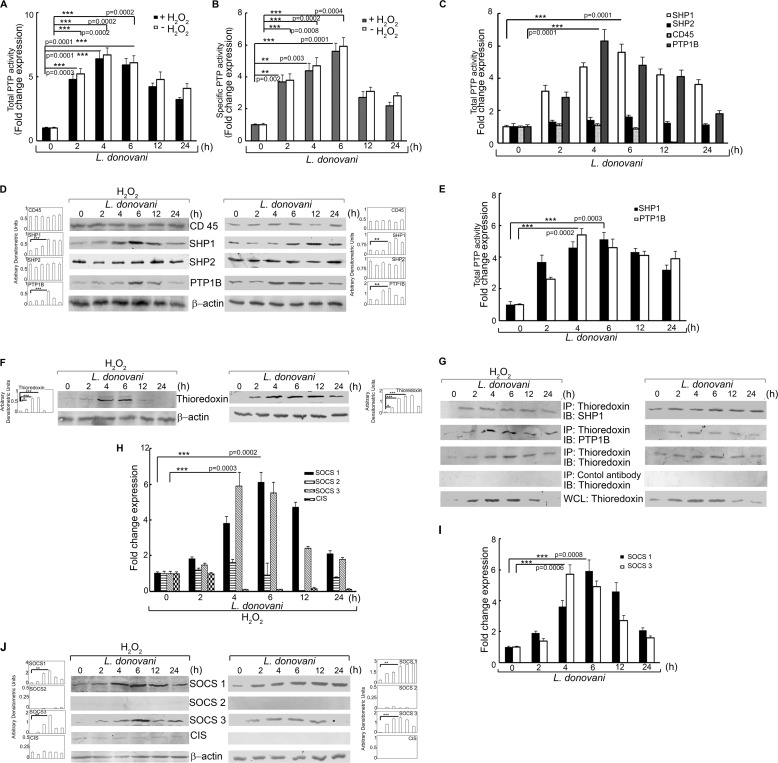
Effect of L. donovani infection on PTP activity, thioredoxin, and SOCS expression. Macrophages were infected with L. donovani for the indicated time periods. One group of infected macrophages from each time point was subjected to H2O2 treatment for 1 h. A and B, total and specific PTP activities were evaluated by the capacity of cell lysates to hydrolyze pNPP (A) or a synthetic tyrosine phosphopeptide (B). Absorbance values were taken at 405 and 620 nm, respectively. C and E, activity of the indicated PTPs were determined by the capacity of immunoprecipitated samples to hydrolyze pNPP in the presence (C) and absence (E) of H2O2. Results are expressed as the relative increase (n-fold) over PTP activity in control cells. D and F, cells were processed as above and then subjected to Western blotting with respective antibodies for various PTPs (D) and thioredoxin (F). G, cells processed as above were immunoprecipitated with anti-thioredoxin antibody followed by immunoblotting with the indicated antibodies. 30 μg of each sample was loaded as a whole cell lysate input control. H–J, expression of various SOCS proteins was determined at mRNA levels in the presence (H) and absence (I) of H2O2 and protein level in the presence (J, left panel) and absence (J, right panel) of H2O2. IP, immunoprecipitation using the indicated antibody; IB, immunoblot analysis using the indicated antibody; WCL, whole cell lysate. Results are representative of three individual experiments, and the error bars represent mean ± S.D. (n = 3). *, p < 0.05; **, p < 0.01; ***, p < 0.001 by Student's t test.

