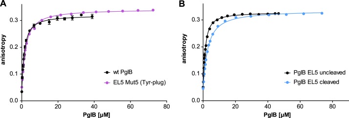
FIGURE 4.

Peptide binding of different EL5 mutants quantified by fluorescence anisotropy. Purified PglB was titrated into a solution containing 1 μm fluorescently labeled peptide variant (containing the sequon DQNAT) and 10 mm MnCl2. Data points reflect the mean of 20 measurements of the same sample. Error bars, S.D. A, binding curve of PglB with replacement of the essential EL5 Tyr-plug sequence element (mutant 5, violet curve) compared with WT PglB (black curve, as presented in Ref. 13). B, binding curve of PglB with a cleavable EL5 (mutant 8 as presented in Fig. 6). The black curve represents PglB with an intact EL5 (uncleaved), whereas the blue curve results from PglB with cleaved EL5.
