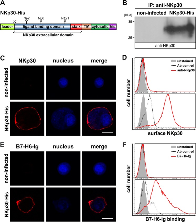
FIGURE 1.
Expression of functional NKp30 in insect cells. A, domain organization of NKp30-His. TM, transmembrane domain; his, decahistidine tag; N42, N68, and N121, acceptor sites for N-linked glycosylation; scissor, cleavage site for leader sequence. B, immunoblot after immunoprecipitation (IP) of NKp30 (anti-NKp30) from lysates of NKp30-His infected or non-infected High Five insect cells. C and D, decoration of non-infected and NKp30-His infected High Five insect cells with an anti-NKp30 antibody to analyze NKp30 surface expression by CLSM (red, anti-NKp30; blue, DAPI; the bar corresponds to 20 μm) and flow cytometry (solid gray, unstained; dashed line, antibody control; red line, anti-NKp30). E and F, B7-H6-Ig fusion protein binding to non-infected and NKp30-His infected High Five insect cells was analyzed by CLSM (red, B7-H6-Ig fusion protein; blue, DAPI; the bar corresponds to 20 μm) and flow cytometry (solid gray, unstained; dashed line, antibody control; red line, B7-H6-Ig fusion protein).

