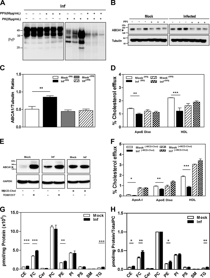
FIGURE 5.
Reversal of the effects of prion infection (Inf). A, cells were treated with PPS (50 μg/ml) or vehicle continuously for 8 days. Western blots of cell lysate with and without PK treatment are shown after development with anti-PrP antibody. B, ABCA1 protein abundance was assessed by Western blotting comparing pre- and post-PPS treatment. C, densitometric quantification of ABCA1 protein abundance before and after treatment with PPS in mock- and prion-infected GT1-7 cells. **, p < 0.01. D, cholesterol efflux to apoE discs (15 μg/ml) and HDL (40 μg/ml) were measured in mock and infected GT1-7 cells, treated and untreated with PPS. **, p < 0.01; ***, p < 0.001. E, ABCA1 protein abundance after prion-infected and mock GT1-7 cells were loaded with cholesterol by incubating with 5 mm cholesterol-loaded methyl-cyclodextrin (MβCD-Chol). F, cholesterol efflux in mock- and prion-infected GT1-7 cells, incubated with or without cholesterol loaded methyl-cyclodextrin. *, p < 0.05; **, p < 0.01; ***, p < 0.001. G and H, lipidomic analysis for mock- and prion-infected GT1-7 cells; absolute values (G) or normalized to PC content (H). CE, cholesterol esters; FC, free cholesterol; Cer, ceramides; PC, phosphatidylcholine; PI, phosphatidylinositol; PS, phosphatidylserine; SM, sphingomyelin; TG, triglycerides.**, p < 0.01; ***, p < 0.001.

