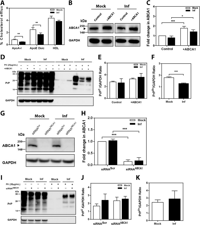
FIGURE 7.
Effect of ABCA1 overexpression and silencing on PrPSc formation in 3T3 cells. A, cholesterol efflux to apoA-I (30 μg/ml), apoE discs (15 μg/ml), and HDL (40 μg/ml) from mock- or prion-infected 3T3 cells; cells were activated with 4 μm LXR agonist (mean ± S. D., n = 3). Inf, infected. **, p < 0.01 versus mock-infected cells. B, Western blot analysis of ABCA1 abundance in 3T3 cells after transient transfection with mock (control) and human ABCA1 plasmid (+ABCA1). C, relative change of ABCA1 abundance in mock- and prion-infected 3T3 cells after transfection with mock and ABCA1 plasmid. ABCA1 abundance was expressed as fold change compared with mock (control) (mean ± S.D., n = 3). *, p < 0.05; ***, p < 0.001. D, Western blot analysis of PrPC and PrPSc in 3T3 cells transiently transfected with mock and human ABCA1 plasmids. E and F, densitometric quantification of PrPC (F) and PrPSc (G) in 3T3 cells transfected with mock and ABCA1 plasmids. (mean ± S.D., n = 3). ***, p < 0.001 versus mock-infected 3T3 cells. G, Western blot analysis of ABCA1 abundance in 3T3 cells after transfection with scrambled (siRNAScr) and mouse ABCA1-specific siRNA (siRNAABCA1, final concentration 20 nm). H, relative protein fold change of ABCA1 in mock-infected and prion-infected 3T3 cells after transfection with siRNAABCA1 or siRNAScr. ABCA1 abundance was expressed as fold change compared with mock-infected cells (mean ± S.D., n = 3). ***, p < 0.001. I, Western blot analysis of PrPC and PrPSc in mock- and prion-infected 3T3 cells transfected with siRNAScr (−) and siRNAABCA1 (+). Densitometric quantification of PrPC (J) and PrPSc (K) in mock- and prion-infected 3T3 cells transfected with siRNAScr and siRNAABCA1.

