
FIGURE 2.
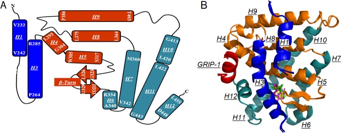
X-ray crystal structures of hRXRα-LBD bound to either Targretin, 9cUAB30, or 9cRA and GRIP-1. A, topology map of holo-hRXRα-LBD showing a three-sandwich helical fold. H4, H5, H8, H9, and the β-sheet (orange) are sandwiched by H1, H2 (missing), and H3 (blue) at one side and H6, H7, H10, and H11 (dark cyan) at the other side. B, overlay of three hRXRα-LBD·GRIP-1·agonist structures. Helices are rendered in the same colors as in A; GRIP-1 is shown in red.