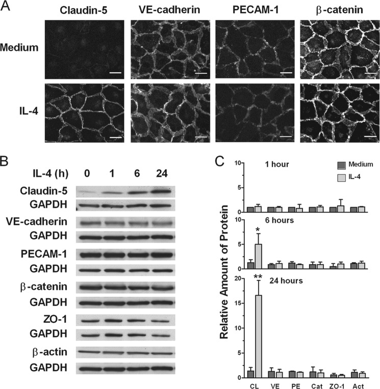
FIGURE 1.
IL-4 induces up-regulation of claudin-5 expression. A, ECs were incubated with medium or 10 ng/ml IL-4 for 40 h, processed for detection of junction proteins claudin-5, VE-catherin, PECAM-1, and β-catenin, and examined by fluorescence microscopy. Results are representative of three independent experiments. Original magnification, ×40. Scale bars, 20 μm. B and C, time-dependent induction of claudin-5 (CL) in comparison with VE-catherin (VE), PECAM-1 (PE), β-catenin (Cat), ZO-1, and β-actin (Act). ECs were incubated with 10 ng/ml IL-4 for 1, 6, or 24 h. Cell extracts were assessed for protein expression using immunoblotting. Bars represent ratios of band density of indicated protein to that of GAPDH loading control relative to time 0 and are means ± S.E. of three experiments. IL-4-treated versus medium alone (*, p = 0.05 and **, p < 0.001). Immunoblots are representative of three independent experiments.

