FIGURE 5.

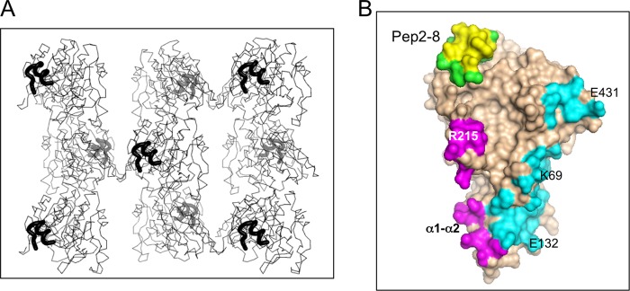
X-ray structure of Pep2-8·PCSK9-ΔCRD; crystal packing contacts. A, Cα traces of Pep2-8·PCSK9-ΔCRD are viewed along the crystal c axis (also the longer axis of the molecule), with Pep2-8 shown as a thick black line. Viewed this way, opposite ends (Pep2-8 binding site and prodomain) alternate foreground/background across and down the figure. B, the 4-Å contact zones from crystal neighbors are shown colored according to different neighboring molecules. Pep2-8 provides an intermolecular contact (yellow) but is otherwise colored green. The neighboring molecule providing the contact colored yellow is omitted from A for clarity.
