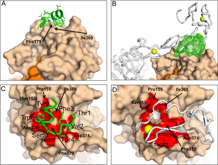
FIGURE 6.
Crystal structure of the Pep2-8·PCSK9-ΔCRD complex. A and B, Pep2-8 binds the catalytic domain of PCSK9-ΔCRD at a region also used by the LDL receptor for binding to PCSK9. A, Pep2-8 (green) is a 13-amino acid peptide composed of a β-strand followed by an α-helix that presents mostly aromatic side chains to the PCSK9 surface (beige). The orange surface is the prodomain of PCSK9. B, the LDL receptor from Protein Data Bank entry 3P5C (gray) is shown with the Pep2-8·PCSK9-ΔCRD complex, after using the catalytic domains for superpositioning. Other colors are as for A, and Pep2-8 is shown as a green mesh. Note that elements of the LDL receptor are inside the Pep2-8 mesh. The yellow spheres represent Ca+2 ions associated with LDL receptor. C and D, close correspondence between PCSK9 surfaces contacted by Pep2-8 and the EGF(A) domain of LDL receptor. C, colored red is the PCSK9 surface (beige) within 4 Å of Pep2-8 (green). Pep2-8 side chains within 4 Å of PCSK9 are included as sticks and are labeled (large type). PCSK9 side chains are labeled in small type. D, colored red is the PCSK9 surface (beige) within 4 Å of EGF(A) (gray), from Protein Data Bank entry 3BPS. The yellow sphere represents a Ca+2 ion as part of the EGF(A) structure. The PCSK9 molecules are displayed in the same orientation after superposition, and selected PCSK9 residues are labeled.

