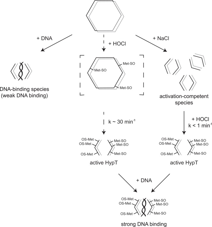
FIGURE 7.
Model of HypT activation. Purified HypT is present as a dodecamer and depicted as two hexamer rings stacked on top of each other. Left, in the presence of DNA, dodecamers dissociate into dimers and tetramers, generating the DNA-binding species with weak DNA-binding activity. Middle, HOCl treatment of dodecamers leads to oxidation of methionine residues and concomitant dissociation of dodecamers into tetramers. This reaction proceeds fairly slowly (k ∼ 30 min−1) and is completed within 1 h. As a result, active HypT tetramers are formed. Right, dissociation of dodecamers into tetramers can be triggered by additives such as NaCl, thus generating activation-competent tetramers. Subsequent treatment with HOCl quickly forms active HypT (k < 1 min−1). Active HypT tetramers show strong DNA-binding activity.

