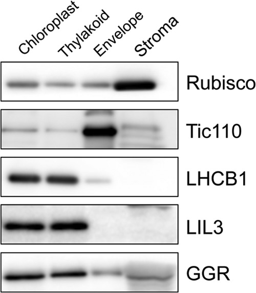
FIGURE 3.

Suborganellar localization of LIL3 and GGR. Chloroplasts were isolated from 4-week-old WT leaves and fractionated into stroma, envelope membranes, and thylakoid membranes. 10 μg of protein was loaded per lane for the analyses with anti-LIL3 and anti-GGR antibodies. 2 μg of protein was loaded per lane for the analyses with the other antibodies. Anti-Rubisco antibody (stroma marker), anti-Tic110 antibody (envelope marker), anti-Lhcb1 antibody (thylakoid marker), anti-LIL3 antibody, and anti-GGR antibody were incubated with protein blots and subsequently detected with a Western Lightning Plus enhanced chemiluminescence kit (PerkinElmer Life Sciences).
