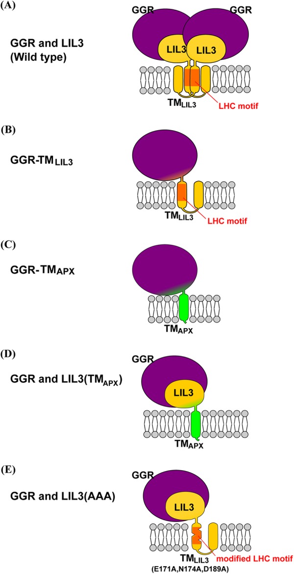
FIGURE 4.

Schematic of the postulated structure of the modified GGR or LIL3 protein. The depicted stoichiometries of each subunit are hypothetical. The large globular structure colored in purple represents GGR, and the small globular structure colored in yellow depicts the N-terminal domain of LIL3. Yellow or green cylindrical structures represent TMLIL3 and TMAPX, respectively. The LHC motif in TMLIL3 is shown in orange. A, postulated structure of the native LIL3-GGR complex. The structure is shown as a dimer, but it could be a trimer or other type of oligomer. B, postulated structure of GGR-TMLIL3. C, postulated structure of GGR-TMAPX. D, structure of the postulated GGR-LIL3(TMAPX) complex. E, postulated structure of the LIL3(AAA)-GGR complex, which appears to be much smaller than the native LIL3-GGR complexes.
