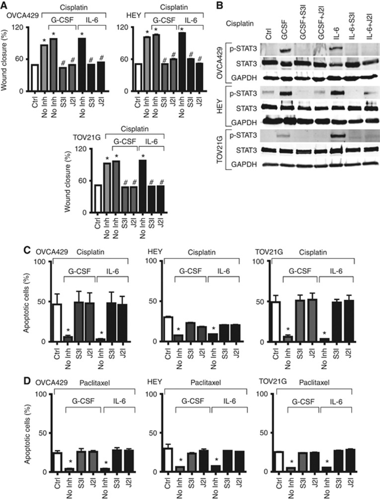
Figure 6.
Effects of chemotherapeutic agents on cell survival and migration. (A) Wound-healing assay. Indicated cells were treated with cisplatin alone, or with G-CSF and IL-6 treatment, in the presence or absence of inhibitors for STAT3 (S3I) and JAK2 (J2I). Graph shows the percentage wound closure, expressed as mean+s.e.m. (*P<0.05 compared with control, #P<0.005 compared with cisplatin). (B) Induction of STAT3 activity. Cell lysates were prepared from OVCA429, HEY and TOV21G cells treated with cisplatin (Cis) as indicated, and subjected to western blot analysis with anti-phospho-STAT3, anti-STAT3 and anti-GAPDH. (C–D) Apoptosis assay. Indicated cells were treated with 12.5 mg ml−1 cisplatin (C) or paclitaxel (D), or with G-CSF and IL-6 treatment, in the presence or absence of inhibitors for STAT3 and JAK2. Graph shows the percentage of apoptotic cells, expressed as mean+s.e.m. (*P<0.005 compared with chemotherapeutic agent). For TOV21G cells, cisplatin was increased to 20 μg ml−1 in order to achieve significant levels of apoptosis.

