Figure 4.
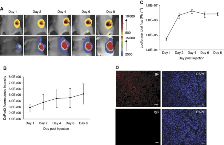
Dual imaging of host cellular proliferation and transcriptional activities of YE-PC8 viruses. (A) ΔGli36-DsRed2 tumours were established in the right flank of SCID mice that were intratumourally injected with an equal amount of YE-PC8 viruses (2 × 10⁵ PFU) directly into the tumours. The DsRed2 fluorescence images were acquired as an indication of tumour growth (represented by top panel). Luciferase activities mediated by YE-PC8 in tumour tissues were imaged at indicated time points after vial injection (represented by the bottom panel). Corresponding quantification of (B) DsRed2 fluorescence signals and (C) luciferase signals. Data were presented as mean±s.e.m., n=4. (D) Tumour sections removed at day 4 post viral administrations and incubated with an anti-gC HSV-1 polyclonal antibody and isotypic control. Scale bar=50 μm.

