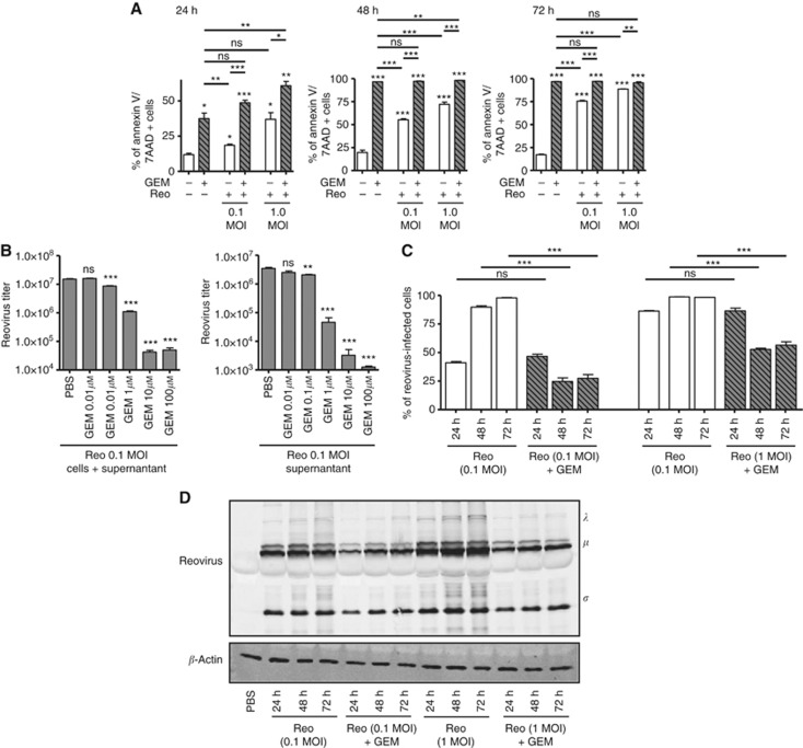
Figure 2.
Increased gemcitabine-induced cell death negatively affects the spread and propagation of reovirus in vitro. MOSE ID8 cells were infected in vitro with 0.1 or 1 MOI of Reo in the presence or absence of 1 μM of gemcitabine and then harvested at 24, 48, and 72 h, stained with annexin-V/7-AAD (detection of apoptotic cells) (A). (B) ID8 cells were infected with Reovirus (0.1 MOI) and treated with various GEM concentrations (0.01, 0.1, 1, 10, or 100 μM). Next, the intracellular and extracellular fractions were collected after 24 h and assessed by standard plaque assay to quantify viral titers (PFU ml−1). (C) Cumulative data on intracellular staining of MOSE ID8 cells with anti-reovirus antibodies to visualise reovirus-infected cells are illustrated. The cumulative data for all conditions tested as noted. The asterisks shown above the horizontal lines display the P-values obtained through comparison between Reo and Reo+GEM groups at the respective time points. Asterisks shown immediately on top of the bars represent the P-values obtained by comparing the respective GEM-treated group against PBS control. Statistical analysis was performed with two-tailed, Student's t-test with 95% CI; ns=P>0.05; *P⩽0.05; **P⩽0.01; ***P⩽0.001. Error bars are defined as mean+s.d. (D) Abundance of reovirus protein in the ID8 cells collected after 24, 48, and 72 h were analysed by western blot. Data are representative of three independent experiments.

