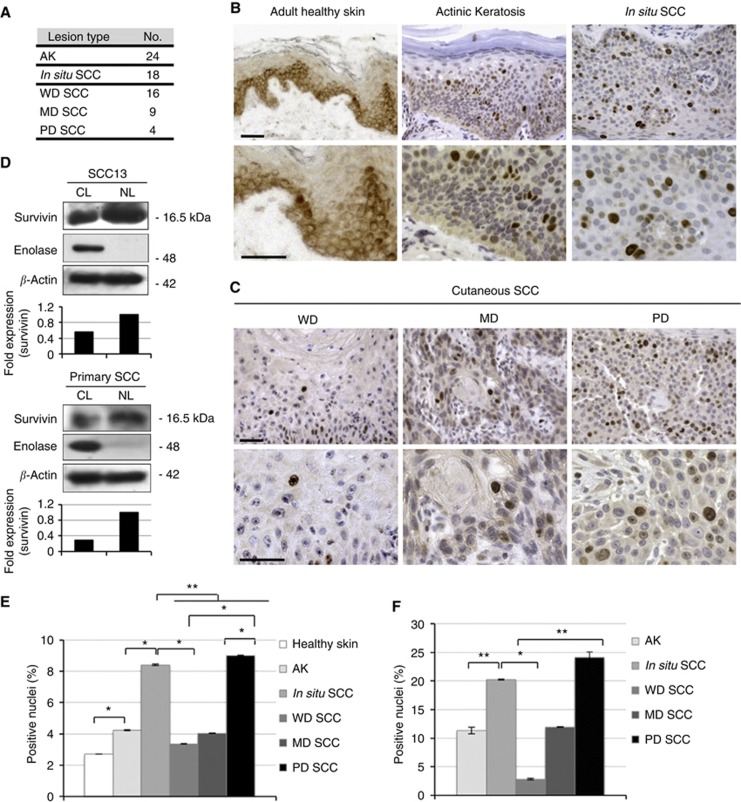
Figure 3.
Expression of N-surv in AK, in situ SCC and cSCC. (A) Number of cases of AK, in situ SCC and cSCC analysed for survivin expression by immunohistochemistry. Cutaneous SCC are divided into WD, MD and PD tumours (see Materials and Methods). (B) Representative immunohistochemical staining of adult healthy skin, AK and in situ SCC for survivin expression (bars=70 μm). (C) Representative immunohistochemical staining of WD, MD and PD cSCC for survivin expression (bars=70 μm). (D) Squamous cell carcinoma 13 cells and total keratinocytes from primary cutaneous SCC were isolated as described in Materials and Methods. NL and CL were obtained and western blot analysis was performed. Enolase was used as a positive control for cytosolic extracts, whereas β-actin was used as a loading control. Band intensity was quantified using Image J software. (E) Quantification of N-surv-positive cells in lesional and healthy skin by using a ‘whole-lesion evaluation' approach (Method I, described in Materials and Methods). *P<0.05; **P<0.01. (F) Quantification of N-surv-positive cells in lesional skin by evaluating areas of the lesions in which survivin staining is positive (Method II, described in Materials and Methods). *P<0.05; **P<0.01.

