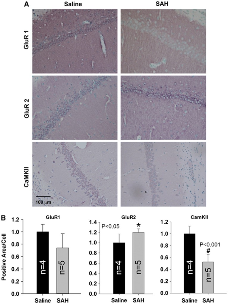
Figure 3.
Immunohistological staining of GluR1, GluR2, and CaMK-II. Representative images are shown in panel A, decreased signal of GluR1 and CaMK-II staining, but increased signal of GluR2 staining was seen in subarachnoid hemorrhage (SAH) group as compared with that in naive or saline controls. Quantified data are shown in panel B. Data are mean±s.d. All animals were killed 6 days after surgery. *P<0.05, #P<0.001.

