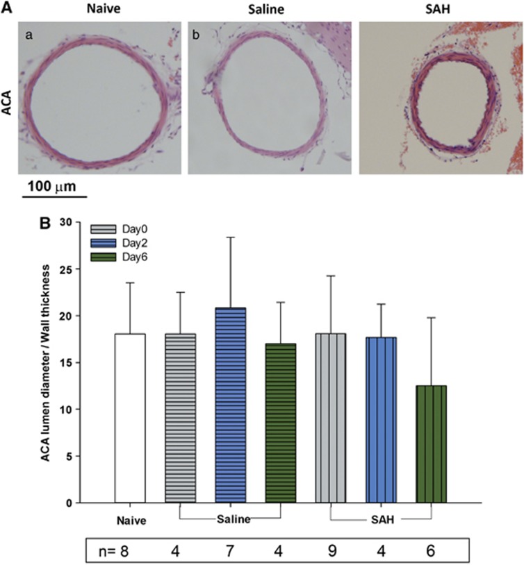
Figure 6.
Vasospasm of anterior cerebral artery (ACA). (A) Representative images showing ACA from different groups of animals. (B) Quantification of lumen diameter/wall thickness ratio in all animal groups. There is no statistical significant difference among the groups (P⩾0.05 analysis of variance). However, there was a trend toward vasospasm in subarachnoid hemorrhage (SAH) day 6 animals (P=0.065 as compared with naive controls). Data are mean±s.d. All animals were killed 6 days after surgery.

