
Figure 1. Structure of the LMO2 in complex with the anti-LMO2 VH#576.
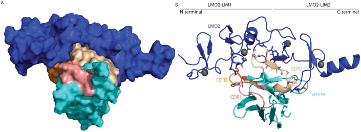
The crystal structure of the dimeric complex of LMO2 and anti-LMO2 VH is shown either in space filling (A) or ribbon form (B). In both, the LMO2 protein is shown in blue and the VH framework region in cyan with CDR regions one, two and three highlighted in salmon, orange and cream respectively. In panel B, the zinc atoms are shown as grey spheres and sticks are used to represent residues involved in inter-molecular hydrogen bonds with oxygen and nitrogen atoms coloured red and blue respectively. For VH#576, there is one residue of CDR one (His31) forming a hydrogen bond, one in CDR two (Ser57) and four in CDR3 (Ser103, Glu105, Thr107 and Trp110).