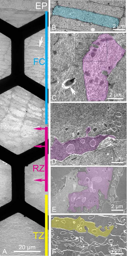
Figure 2. Thin section TEM montage from the epithelium to a depth of about 150 µm recorded in the equatorial plane from an adjacent region in the same sample as in Fig. 1.
A Overview montage showing four distinct regions: epithelium (EP), classical fiber cells (FC; cyan, 80 µm thick), remodeling zone (RZ; magenta, 40 µm thick), and transitional zone (TZ; yellow, >500 µm thick). Many cells in FC contain nuclei even though only one is shown (arrow). Arrows on the magenta bar represent the depth from the epithelium where enlargements are made. Black bars are visible from the 300 mesh hexagonal support grid. B. Higher magnification of a classical fiber cell indicates the 2 µm × 10 µm flattened hexagonal shape with smooth broad faces interrupted by only a few small edge processes (small circular profiles). C. Beginning of RZ (upper magenta arrow in A) reveals numerous intercellular projections that look like ball-and-socket interlocking devices (two remain connected in the highlighted cell). The bulbous ends stain darkly against a light and textured cytoplasm. The boxed region is enlarged in Fig. 3. A vesicle containing heterogeneous debris is probably a secondary lysosome (arrow). D. The middle of the RZ (middle magenta arrow in A) is characterized by cells of irregular shape with many interdigitating processes of varying size and staining density (highlighted cell). E. Deep in the RZ (lower magenta arrow in A) cells are still very irregular in shape and internal projections are variable. In some cells projections are nearly 2 µm in diameter and irregular in outline with smooth interiors similar to the adjacent cytoplasm (highlighted cell). The narrow cell to the right has the majority of the cytoplasm filled with projections whereas other cells have very few. F. Beginning of the TZ has cells, which are more uniform in staining density and texture with fewer projections. Although the cell shape is still irregular, radial cell columns can be identified and early stages of compaction and undulating membranes can be recognized (highlighted cell).

