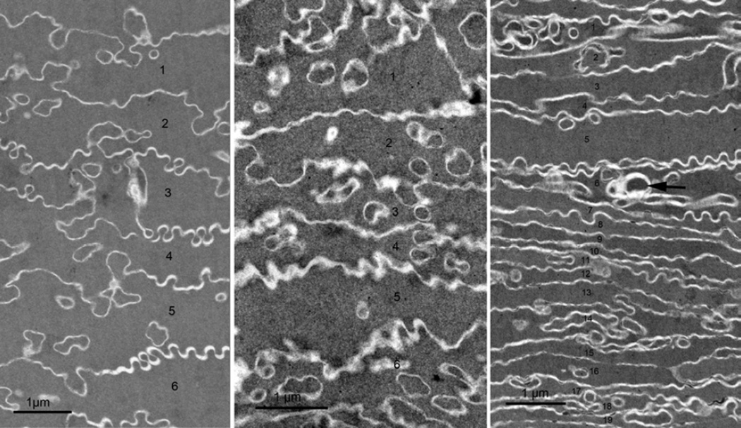
Figure 5. Fiber cell compaction as a function of lens age.
TEM images were recorded at about 700 µm from the capsule near the adult nucleus where all the cells have irregular shapes. A. Very little compaction was noted in the 22 y.o. donor lens. The six cells labeled 1–6 have an average thickness of approximately 1.1 µm (compared to 2 µm in the outer cortex in region FC in Fig. 1A). B. Somewhat greater compaction is seen in the 55 y.o. donor lens, here about 0.8 µm average cell thickness for six cells 1–6. C. In the 92 y.o. donor lens, the cell width is variable with the average for this field of 19 cells (labeled 1–19) is about 0.4 µm. Note also that focal defect structures (arrow) are more common in the older lens.

