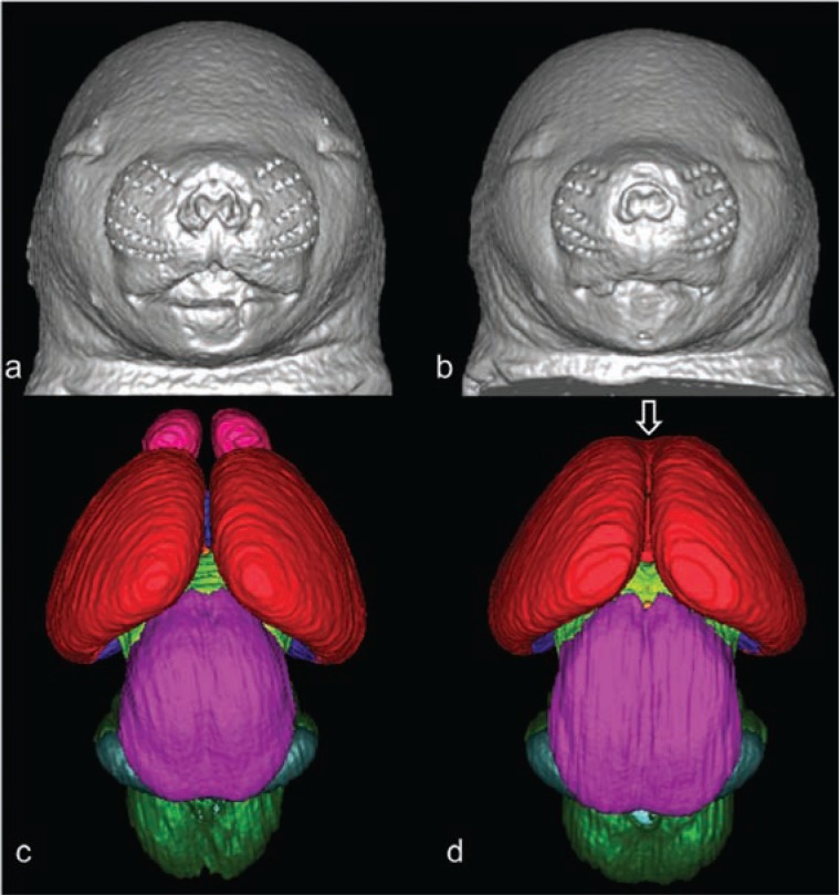
Figure 3.
3D reconstruction of the faces and brains of mice at 17 days of gestation. Control mice are shown in a and c, mice exposed to alcohol are shown in b and d. Mouse fetuses in b and d illustrate dysmorphology resulting from exposure to alcohol at 7 days of gestation. Compared to the control (a), the ethanol-exposed fetus (b) has a smaller head size, a small nose, and an elongated/abnormal philtral portion of the upper lip. These facial features are characteristic of fetal alcohol syndrome. The brain of the ethanol-exposed animal also is dysmorphic (d); the olfactory bulbs are absent and the cerebral hemispheres are united rostrally (open arrow). Color codes: red=cerebral cortex, pink=olfactory bulbs, magenta=mesencephalon, light green=diencephalon, dark green=pons and medulla, teal=cerebellum.
SOURCE: O’Leary-Moore, S.K.; Parnell, S.E.; Godin, E.A.; and Sulik, K.K. Magnetic resonance-based studies of FASD in animal models, Alcohol Research & Health, in press. Modified from Godin, E.A.; O’Leary-Moore, S.K.; Khan, A.A.; et al. Magnetic resonance microscopy defines ethanol-induced brain abnormalities in prenatal mice: effects of acute insult on gestational day 7. Alcoholism: Clinical and Experimental Research 34(1):98–111, 2010. PMID: 19860813

