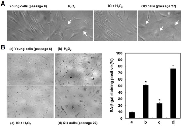
Fig. 3. Effect of I. obliquus on hydrogen peroxide-induced premature senescence in human dermal fibroblasts. The cells were treated with 500 μM hydrogen peroxide for 2 h in the presence or absence of I. obliquus extract (25 μg/ml) and then recovered for 7 days with fresh medium. (A) Morphological observation of cells was detected by optical microscopy. Magnification: 200-fold. Arrows indicate changed morphology, such as flattened and irregular cell morphology or enlarged cell size according to premature senescence. (B) Representative micrographs of young cells (passage 6), 500 μM hydrogen peroxide- induced premature senescent cells, I. obliquus-treated cells, and old cells (passage 27) after SA-β-gal staining. Magnification: 100-fold. The percentage of SA-β-gal positively stained cells was quantified as described in Section 2. Data represents the mean ± SE of three independent experiments. Significant differences were compared with the control at *p < 0.05 by Student’s t-test.

