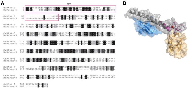
Fig. 3. (A) Sequence alignment between C. elegans RABX-5 and bovine Rabex-5. The region which is disrupted in the C. elegans rabx-5(tm1512) mutant is shown as a purple box. Black inverted triangles refer to the key residues in recognizing ubiquitin in bovine Rabex-5. (B) Homology modeling of C. elegans RABX-5 A20 zinc finger domain using SwissModel Server. C. elegans RABX-5 is colored in purple with zinc ion in red. Bovine Rabex-5 spanning A20 zinc finger and motif-interacting with ubiquitin (MIU) is depicted in gray with two bound ubiquitin molecules in blue and yellow.