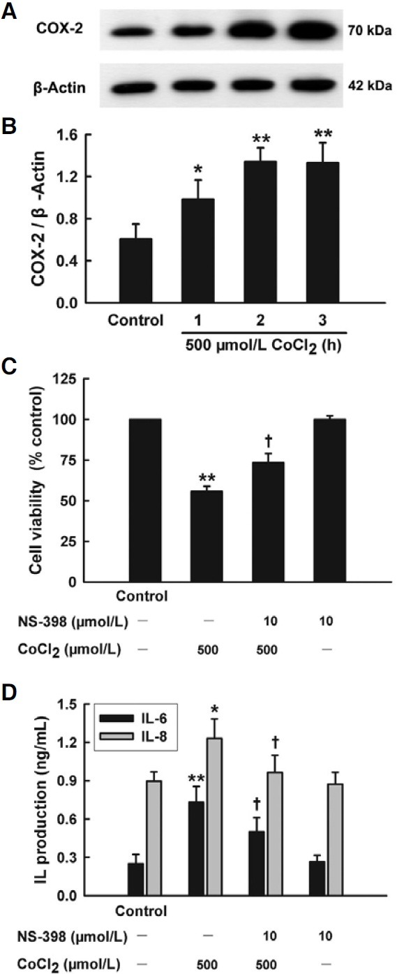
Fig. 3. Roles of COX-2 in cytotoxicity and inflammation induced by CoCl2 in HaCaT cells. (A) HaCaT cells were treated with 500 μmol/L CoCl2 for the indicated time (1-3 h). COX-2 expression was detected by Western blot assay. (B) Densitometric analysis results from A. (C, D) HaCaT cells were treated with 500 μmol/L CoCl2 for 24 h in the presence or absence of pretreatment with 10 μmol/L NS-398 for 30 min. CCK-8 assay was applied to measure cell viability (C). ELISA was used to detect the levels of IL-6 and IL-8 in cell culture medium (D). Data are the mean ± SE (n = 3). *P < 0.05, **P < 0.01 compared with control group. + P < 0.05 compared with CoCl2 treatment group.

