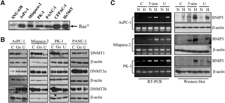
Fig. 3. Effect of U0126 treatment on the expression of DNMT and BNIP3. (A) The 30 μg of total cell extract of each cell line was incubated with a Raf-1-RBD GST fusion protein in glutathione agarose. The captured protein was eluted, and analyzed by Western blotting using anti-Ras monoclonal antibody. (B) The indicated pancreatic cancer cell lines were treated with vehicle only (C), 10 μM U0126 (U) or 5 μM Go6976 (Go) for 6 days. Cell lysates were analyzed for expression of DNMTs by immunoblotting with antibodies against DNMT1, DNMT3a, and DNMT3b. Data are representative of three independent experiments. β-actin was used as an internal control. (C) Cells were treated with vehicle only (C), 1 μM 5-aza-2-deoxycytidine (5-aza) or 10 μM U0126 (U) for 6 days before exposure to hypoxic condition for a further 48 h. BNIP3 mRNA (left panel) and protein (right panel) levels were examined by RT-PCR and immunoblotting, respectively. The β-actin was used as a control. N, normoxia, H, hypoxia.