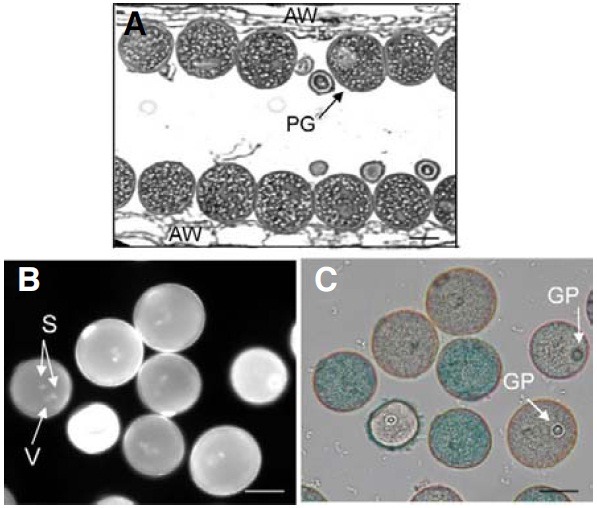
Fig. 4. Transmission electron microscopy analysis of OsImpβ1/osimpβ1-1 anther. Longitudinal sections of OsImpβ1/osimpβ1-1 anther at mature stage (A). Pollen grains from the OsImpβ1/osimpβ1-1 anther at mature stage were stained with DAPI (B) and GUS solutions (C). Stained pollen grains were monitored with a fluorescence microscope (B) and a bright-field microscope (C). AW, anther wall; GP, germination pore; PG, pollen grain; S, sperm nuclei; V, vegetative nucleus. Bars = 50 μm.

