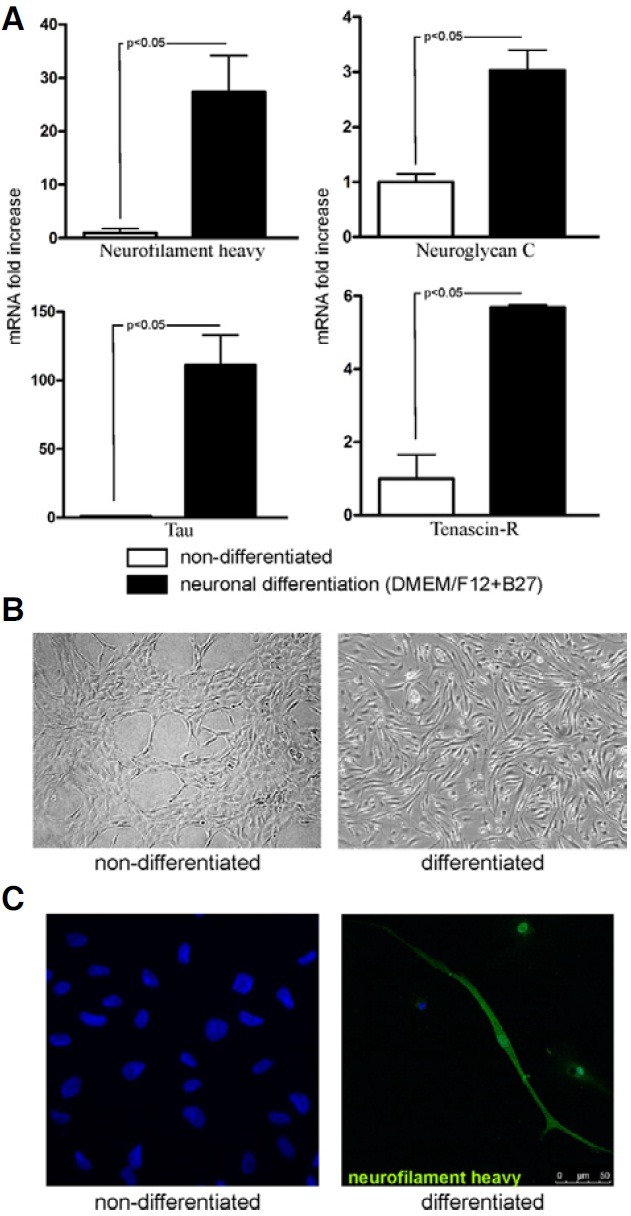
Fig. 5. Neuronal differentiation of expanded MSC. (A) Expression of neuroglycan C, neurofilament heavy, tau and tenascin-R evaluated in expanded MSC after neuronal differentiation by real-time RTPCR, when compared to non-differentiated MSC. The expression levels of neuronal markers were evaluated in two independent representative differentiation experiments. Results are presented as mean ± SEM. (B) Morphology of MSC undergoing neuronal differentiation (magnification 100×). The morphology of cells changed during culture resulting in elongated, needle-like phenotype of cells after one week of culture in neuronal conditions. (C) Confocal microscopy analysis confirmed the neurofilament heavy protein expression in cells acquiring the neuron-like morphology after neuronal differentiation.