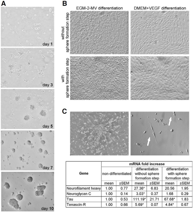
Fig. 6. Sphere formation and differenttiation of sphere-derived cells. (A) Process of sphere/cluster formation. At 24 h after seeding few cells adhered to the poli-D-lysine coated dish (day 1). The cells migrated during next 3-4 days forming cellular aggregates initiating spheres (day 3). Rapid proliferation of these cells resulted in increased size of clusters (day 5). Finally, around one week after seeding some of the large clusters spontaneously detached from the culture dish and grew into floating round spheres (day 7). When the culture was prolonged the fusion of floating spheres was observed (day 10) (magnification 100×). (B) Morphology of expanded MSC without or with sphere formation step following endothelial differentiation (magnification 100×). The cells were culture in EGM- 2-MV defined medium or DMEM supplemented with VEGF. (C) The neuronal differentiation of MSC with and without the sphere formation step. The cells acquired the elongated, needle-like bipolar morphology (magnification 100×). Proliferating cells are indicated by arrows (magnification 200×). Real-time RTPCR analysis showed the changes in expression of neuronal markers on mRNA level in differentiated MSC (without and with sphere formation step) when compared to non-differentiated cells. The expression levels of indicated markers were evaluated in two representative differentiation experiments. Results are presented as mean ± SEM. (*) p < 0.05 vs. non-differentiated cells.

