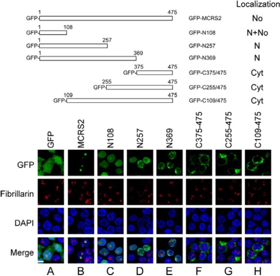
Fig. 1. Map and distribution of MCRS2 mutants. 293T cells were transfected with plasmids that express GFP, GFP-MCRS2, GFPN108, GFP-N257, GFP-N369, GFP-C375/ 475, GFP-C255/475, and GFP-C109/475. At 18 h after transfection, proteins in the cell were stained with anti-fibrillarin antibody and Alexa 594-conjugated goat anti-mouse IgG antibody to reveal the nucleolus (No). The nucleus (N) was stained using DAPI. Cyt, cytoplasm; bar, 10 micrometer.

