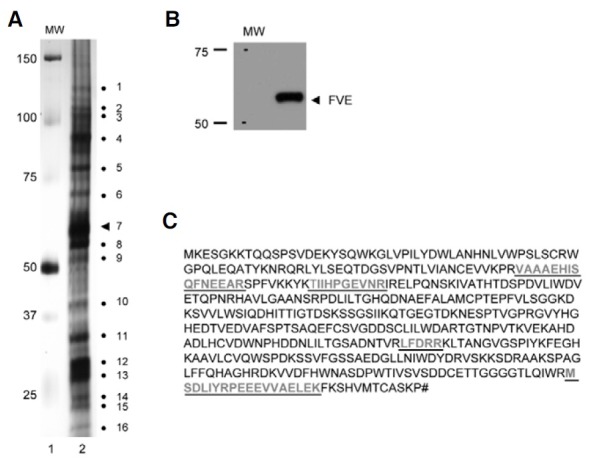
Fig. 5. SDS-PAGE analysis of proteins associated in the FVE complex. (A) SDS-PAGE analysis of the fractions corresponding to the FVE complexes after immunoprecipitation with FLAG antibody. The FVE protein complexes collected by gel-filtration chromatography from 44 to 52 fractions were incubated with 50 μl of anti-FLAG M2 affinity resin, precipitated, and eluted with the 3× FLAG peptide. The elutes (lane 2) were then loaded on 12% SDS-PAGE and silver-stained. The numbers to the left are the sizes of the molecular weight marker proteins (lane 1). Numbers to the right show the apparent molecular weights of the polypeptides detected by silver staining in decreasing order (lane 2). The triangle numbered 7 indicates FVE. (B) Immunoblot analysis of the FVE complexes immunoprecipitated with FLAG antibody. The immunoprecipitated FVE protein complexes were loaded onto 12% SDS-PAGE and subjected to immunoblot analysis with HA antibody. (C) Peptide sequence of FVE detected by MALDI-MS. The FVE complexes immunoprecipitated with FLAG were used for 1-D gel electrophoresis. The number 7 band (A) was identified as FVE. Underlined bold letters indicate the peptides detected by MALDI-MS. The matched peptides represent 11% amino acid sequence coverage.

